Home » Without Label » Smooth Muscle Diagram Labeled - Skeletal Muscle Organization / Pericytes allow smooth muscle cells to regenerate and repair much more readily than skeletal and cardiac muscle tissue.
Smooth Muscle Diagram Labeled - Skeletal Muscle Organization / Pericytes allow smooth muscle cells to regenerate and repair much more readily than skeletal and cardiac muscle tissue.
Smooth Muscle Diagram Labeled - Skeletal Muscle Organization / Pericytes allow smooth muscle cells to regenerate and repair much more readily than skeletal and cardiac muscle tissue.. Drag the labels onto the diagram to label the steps of smooth muscle activation and deactivation. This might sound like a strange question, right? Related posts of smooth muscle labelled diagram muscle anatomy back. Smooth muscle fibers are often found forming sheets of tissue and function in a coordinated fashion due to the presence of gap junctions between the cells. Smooth muscle tissue, unlike striated muscle, contracts slowly and automatically.
You will also get the identification points of skeletal muscle histology slide with a little description here in this guide. Muscle anatomy back 12 photos of the muscle anatomy back back muscle anatomy images, back muscle anatomy of the human body, back pain muscle anatomy, muscle anatomy lower back, posterior back muscle anatomy, human muscles, back muscle anatomy images, back muscle anatomy of the human body, back pain muscle anatomy, muscle. Name the tough connective tissue cord that serves to attach a muscle to a bone. 1024x840 draw a labelled diagram of a smooth muscle diagram of smooth. Nine characteristics of muscle tissue are listed below and on page 104.
Muscular Tissue Class 9 Tissues from classnotes.org.in In this video i have shown the simplest way of drawing muscle drawing. Similar to skeletal muscle tissue, cardiac muscle does not regenerate to a great extent. Smooth muscle (textus muscularis levis) smooth muscle is a type of tissue found in the walls of hollow organs, such as the intestines, uterus and stomach. Overview of muscle tissues 1. Which of the labeled layers in the diagram of the arterial wall is composed of a simple squamous epithelium, a basement membrane and a layer of elastic tissue? Smooth muscle contracts under certain stimuli as atp is freed. Smooth muscle is composed of sheets or strands of smooth muscle cells. Structure and composition of muscle meat science.
Identify the muscle tissue type described by choosing the correct response(s) from the key choices.
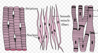
The term smooth muscle refers to the more uniform appearance of this type of muscle tissue. Structure and composition of muscle meat science. They range from about 30 to 200 μm (thousands of times shorter than skeletal muscle fibers), and they produce their own connective tissue, endomysium.although they do not have striations and sarcomeres, smooth muscle fibers do have actin and myosin. Drag the labels onto the diagram to label the steps of smooth muscle activation and deactivation. Skeletal muscles attach to and move bones by contracting and relaxing in response to voluntary messages from the nervous system. Smooth muscle is a type of muscle tissue which is used by various systems to apply pressure to vessels and organs. This diagram shows a few of the cells that can be seen in the stained section below. The cells are spindle shaped, and the nucleus is central. Smooth muscle is a type of tissue found in the walls of hollow organs, such as the intestines, uterus you can also find smooth muscle in the walls of passageways, including arteries and veins. Muscle fibers are organized into bundles supplied by blood vessels and innervated by motor neurons. 1024x840 draw a labelled diagram of a smooth muscle diagram of smooth. Nine characteristics of muscle tissue are listed below and on page 104. These cells have fibers of actin and myosin which run through the cell and are supported by a framework of other proteins.
These cells have fibers of actin and myosin which run through the cell and are supported by a framework of other proteins. I mean, the abs are the muscle. The cells are spindle shaped, and the nucleus is central. 1024x840 draw a labelled diagram of a smooth muscle diagram of smooth. The skeletal muscle fibers are elongated, cylindrical and multinucleated cells whose length may vary in different animals.
I Answer The Following A Draw A Labeled Diagram Of Smooth Muscle Give One Difference Between Yudem And Nh from toppr-doubts-media.s3.amazonaws.com The cells are spindle shaped, and the nucleus is central. Pericytes allow smooth muscle cells to regenerate and repair much more readily than skeletal and cardiac muscle tissue. Its wavelike movements propel things through the bodily system, such as food through. Smooth muscle diagram labeled the muscular system micro and macro anatomy, picture of smooth muscle diagram labeled the muscular system micro and macro anatomy Overview of muscle tissues 1. Which of the labeled layers in the diagram of the arterial wall consists mainly of elastic fibers and smooth muscle fibers? Drag the labels onto the diagram to label the steps of smooth muscle activation and deactivation. You will also get the identification points of skeletal muscle histology slide with a little description here in this guide.
Its wavelike movements propel things through the bodily system, such as food through.
Cardiac s m s a c b. This diagram shows a few of the cells that can be seen in the stained section below. Name the tough connective tissue cord that serves to attach a muscle to a bone. Smooth muscle contracts under certain stimuli as atp is freed. Smooth muscle is made up of cells that contain a single central nucleus. The anatomy of your abdominal muscles. In this video i have shown the simplest way of drawing muscle drawing. In skeletal muscle, a single type of somatic nervous system traverses to muscle, where it stimulates organelle in the muscle cells in order to release calcium. The skeletal muscle fibers are elongated, cylindrical and multinucleated cells whose length may vary in different animals. Smooth muscles are unique in their largely involuntary response, and in their structure. Smooth muscle (textus muscularis levis) smooth muscle is a type of tissue found in the walls of hollow organs, such as the intestines, uterus and stomach. But in actuality there are 4 separate muscles that contribute to your overall abdominal development. Structure and composition of muscle meat science.
In skeletal muscle, a single type of somatic nervous system traverses to muscle, where it stimulates organelle in the muscle cells in order to release calcium. Diagram of smooth muscle cardiac muscle and straited muscle tissue. Skeletal muscles attach to and move bones by contracting and relaxing in response to voluntary messages from the nervous system. Drag the labels onto the diagram to label the steps of smooth muscle activation and deactivation. The skeletal muscle fibers are elongated, cylindrical and multinucleated cells whose length may vary in different animals.
Cbse Ncert Notes Class 9 Biology Tissues from www.examfear.com 1024x840 draw a labelled diagram of a smooth muscle diagram of smooth. Name three types of fiber arrangements seen in skeletal muscle. Smooth muscle tissue can regenerate from a type of stem cell called a pericyte, which is found in some small blood vessels. This might sound like a strange question, right? Smooth muscle makes up the walls of hollow organs, respiratory passageways, and blood vessels. Smooth muscle cells are often rounded at the center and tapered off at the sides. Smooth muscle diagram labeled the muscular system micro and macro anatomy, picture of smooth muscle diagram labeled the muscular system micro and macro anatomy Smooth muscle, muscle that shows no cross stripes under microscopic magnification.
1024x840 draw a labelled diagram of a smooth muscle diagram of smooth.
The skeletal muscle fibers are elongated, cylindrical and multinucleated cells whose length may vary in different animals. I mean, the abs are the muscle. Muscle fibers are organized into bundles supplied by blood vessels and innervated by motor neurons. Which of the labeled layers in the diagram of the arterial wall is composed of a simple squamous epithelium, a basement membrane and a layer of elastic tissue? Smooth muscle cells are often rounded at the center and tapered off at the sides. There are three types of muscle in the body: Smooth muscle is composed of sheets or strands of smooth muscle cells. Smooth muscle tissue, unlike striated muscle, contracts slowly and automatically. Smooth muscle cells are found in the dividers of empty organs, including the stomach, digestion tracts, urinary bladder and uterus, and in the dividers of paths, for example, the supply routes and veins of the circulatory framework, and the tracts of the. Different muscle types in the human body. Smooth muscle fibers are often found forming sheets of tissue and function in a coordinated fashion due to the presence of gap junctions between the cells. Identify the muscle tissue type described by choosing the correct response(s) from the key choices. They range from about 30 to 200 μm (thousands of times shorter than skeletal muscle fibers), and they produce their own connective tissue, endomysium.although they do not have striations and sarcomeres, smooth muscle fibers do have actin and myosin.
In skeletal muscle, a single type of somatic nervous system traverses to muscle, where it stimulates organelle in the muscle cells in order to release calcium smooth muscle diagram. 1024x840 draw a labelled diagram of a smooth muscle diagram of smooth.